43 compound microscope labelling
What is a Compound Microscope? | Microscope World Blog A compound microscope is an upright microscope that uses two sets of lenses (a compound lens system) to obtain higher magnification than a stereo microscope. A compound microscope provides a two-dimensional image, while a stereo microscope provides a three-dimensional image. Compound microscopes typically provide magnification in the range of ... What is a Compound Microscope? - Study.com The compound microscope, also called compound light microscope, is an upright microscope that utilizes two lenses to magnify objects. It gets its name because it uses two lenses added to each ...
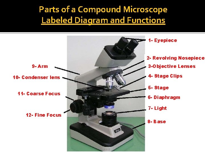
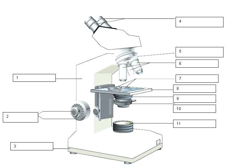
Labeling the Parts of the Microscope | Microscope activity, Science ... Jan 13, 2016 - Free worksheets for labeling parts of the microscope including a worksheet that is blank and one with answers. ... Description Worksheet identifying the parts of the compound light microscope. Answer key: 1. Body tube 2. Revolving nosepiece 3. Low power objective 4. Medium power objective 5. High power objective 6. Stage clips 7.
Compound microscope labelling
Lab Supplies | Lab Consumables | Lab Equipment | Hurst Scientific We supply lab equipment, lab supplies and lab consumables for most scientific disciplines. Browse our wide range of lab and scientific products online. Delivered Australia wide. Microscope Labeling - The Biology Corner Microscope Labeling. Shannan Muskopf May 31, 2018. This simple worksheet pairs with a lesson on the light microscope, where beginning biology students learn the parts of the light microscope and the steps needed to focus a slide under high power. The labeling worksheet could be used as a quiz or as part of direct instruction where students ... 10 Best Compound Microscopes (Summer 2022) - The Complete Guide Celestron Labs designed a premium compound microscope with exceptional durability. It comes with a two-year warranty, but it will last longer if you maintain it right. The product includes two eyepieces, a WP-20x and a WF-10x with a pointer. You can change these eyepieces easily to find your perfect adjustment.
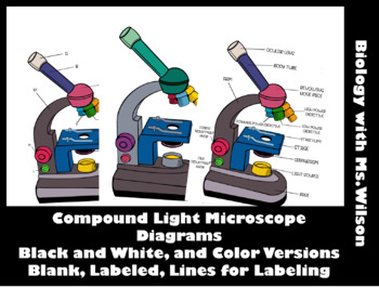
Compound microscope labelling. label parts of a compound microscope - TeachersPayTeachers This is from a set of 3 tiered readings. Students will read a passage about the how to use a compound light microscope. Students will use textual evidence to answer questions and label the different parts of the microscope. It also allows students to gain prior knowledge about the compound microscope. Version A provides the most support for ... Parts of a microscope with functions and labeled diagram - Microbe Notes Head - This is also known as the body. It carries the optical parts in the upper part of the microscope. Base - It acts as microscopes support. It also carries microscopic illuminators. Arms - This is the part connecting the base and to the head and the eyepiece tube to the base of the microscope. Compound Microscope- Definition, Labeled Diagram, Principle, Parts, Uses Alternatively, the magnification of the compound microscope is given by: m = D/ fo * L/fe where, D = Least distance of distinct vision (25 cm) L = Length of the microscope tube fo = Focal length of the objective lens fe = Focal length of the eye-piece lens Parts of a Compound Microscope Eyepiece And Body Tube. Compound Microscope Parts - Labeled Diagram and their Functions The term "compound" refers to the microscope having more than one lens. Basically, compound microscopes generate magnified images through an aligned pair of the objective lens and the ocular lens. In contrast, "simple microscopes" have only one convex lens and function more like glass magnifiers. [In this figure] Two "antique" microscopes played significant roles in the history of biology.
NCERT Class 9 Science Lab Manual – Plant and Animal Tissues Permanent slides of parenchyma tissues, sclerenchyma tissues, straited muscle fibre, nerve cell and compound microscope. Procedure. Place the compound microscope where proper light can be received and reflected on the slide. Place the permanent slides one by one. Observe its structure and draw diagrams. Observations I. Plant tissues PDF Label compound microscope worksheet - Weebly Label compound microscope worksheet Name_____. In paragraph 15, when you concentrating the sample, you should always start with the goal of _____ 16. Only the _____ button should be used when using a high-power target. 17. The type of microscope used in most science classes is the _____ microscope. Phosphotungstic acid - Wikipedia Phosphotungstic acid (PTA) or tungstophosphoric acid (TPA), is a heteropoly acid with the chemical formula H 3 P W 12 O 40].It forms hydrates H 3 [PW 12 O 40]·nH 2 O.It is normally isolated as the n = 24 hydrate but can be desiccated to the hexahydrate (n = 6). Compound Microscope: Parts of Compound Microscope - BYJUS The parts of the compound microscope can be categorized into: Mechanical parts; Optical parts (A) Mechanical Parts of a Compound Microscope. 1. Foot or base. It is a U-shaped structure and supports the entire weight of the compound microscope. 2. Pillar. It is a vertical projection. This stands by resting on the base and supports the stage. 3. Arm
How to draw compound of Microscope easily - step by step Perhaps Bidesh. 42.9K subscribers. I will show you " How to draw compound of microscope easily - step by step " Please watch carefully and try this okay. Thanks for watching..... # ... Compound Light Microscope: Everything You Need to Know A compound light microscope is a type of light microscope that uses a compound lens system, meaning, it operates through two sets of lenses to magnify the image of a specimen. It's an upright microscope that produces a two-dimensional image and has a higher magnification than a stereoscopic microscope. It also goes by a couple of other names ... Soil networks become more connected and take up more carbon … 08.02.2017 · Soil organisms have an important role in aboveground community dynamics and ecosystem functioning in terrestrial ecosystems. However, most studies have considered soil biota as a black box or ... Compound Microscope - Types, Parts, Diagram, Functions and Uses Compound microscope - It has two convex lenses. It is called a compound microscope because it compounds the light as it passes through the lenses to magnify. The image of the object being viewed is enlarged because of the lens near the object. An eyepiece, an additional lens, is where real magnification takes place.
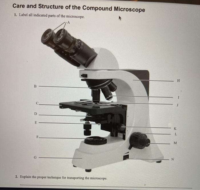
Solved Care and Structure of the Compound Microscope 1 ...
Diagram of a Compound Microscope - Biology Discussion 1. It is noted first that which objective lens is in use on the microscope. 2. Stage micrometer is positioned in such a way that it is in the field of view. 3. The eyepiece is rotated so that the two scales, the eyepiece or ocular scale and the stage micrometer scale, are parallel. 4.

Monday, 12 November 2018Monday, 12 November ppt download
Microscope Labeling Practice Quiz - PurposeGames.com This is an online quiz called Microscope Labeling Practice There is a printable worksheet available for download here so you can take the quiz with pen and paper. From the quiz author Practice labeling a compound microscope. Do you know all the parts? This quiz has tags. Click on the tags below to find other quizzes on the same subject. microscope

Solved Microscope parts/labeling 9 Label the image of a ...
compound microscope parts (labeling) Flashcards | Quizlet compound microscope parts (labeling) Flashcards | Quizlet compound microscope parts (labeling) STUDY Flashcards Learn Write Spell Test PLAY Match Gravity Created by barnettlily Terms in this set (14) eyepiece tube - connects the eyepiece to the objective lens what is 1? nosepiece (turret) - holds and spins the objective lenses what is 2?

File:Microscope diagram.png - Wikimedia Commons
Labelled Diagram of Compound Microscope - Biology Discussion The below mentioned article provides a labelled diagram of compound microscope. Part # 1. The Stand: The stand is made up of a heavy foot which carries a curved inclinable limb or arm bearing the body tube. The foot is generally horse shoe-shaped structure (Fig. 2) which rests on table top or any other surface on which the microscope in kept.

Compound Microscope Parts, Functions, and Labeled Diagram ...
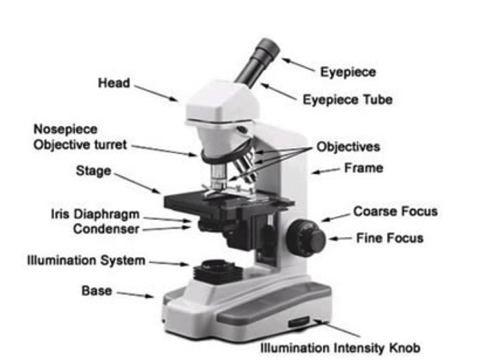
Parts of the Microscope with Labeling (also Free Printouts) Parts of the Microscope with Labeling (also Free Printouts) By Editorial Team March 7, 2022 A microscope is one of the invaluable tools in the laboratory setting. It is used to observe things that cannot be seen by the naked eye. Table of Contents 1. Eyepiece 2. Body tube/Head 3. Turret/Nose piece 4. Objective lenses 5. Knobs (fine and coarse) 6.

Compound Microscope Parts, Functions, and Labeled Diagram ...
Compound Microscope - Diagram (Parts labelled), Principle and Uses What actually is a compound microscope? Also called as binocular microscope or compound light microscope, it is a remarkable magnification tool that employs a combination of lenses to magnify the image of a sample that is not visible to the naked eye. Compound microscopes find most use in cases where the magnification required is of the higher order (40 - 1000x). The magnification effect is achieved using the combination of the objective lens (near the sample) and the ocular lens (within ...

The Parts of a Compound Microscope and How To Handle Them ...
Home Page [ ] Our Company News / Blog : 6th June 2022 Congratulations John! Celebrating 50 years in the Science Industry - what a huge milestone! Our 1300 numbers are no longer active.
The Microscope
PharmaCircle This website uses cookies to help provide you with the best possible online experience. Please read our Terms & Conditions and Privacy Policy for information about ...

This is a common compound microscope. Label its parts from A ...
ProSciTech Laboratory supplies and Lab equipment for Histology, Pathology, Light Microscopy, Electron Microscopy and specialist researchers.
Welcome to Microbiology Lab King Saud University Dept
Label the Skeleton Quiz - PurposeGames.com About this Quiz. This is an online quiz called Label the Skeleton. There is a printable worksheet available for download here so you can take the quiz with pen and paper.. This quiz has tags. Click on the tags below to find other quizzes on the same subject.
Understanding the Compound Microscope Parts and its Functions ...
Labeling the Parts of the Microscope | Microscope World Resources Microscope World explains the parts of the microscope, including a printable worksheet for schools and home. Need Asssistance? 800-942-0528. Microscope Blog ... Labeling the Parts of the Microscope. This activity has been designed for use in homes and schools. Each microscope layout (both blank and the version with answers) are available as PDF ...
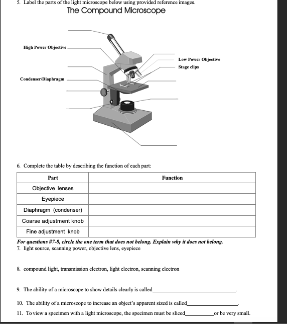
Solved 5. Label the parts of the light microscope below ...
Label the microscope — Science Learning Hub All microscopes share features in common. In this interactive, you can label the different parts of a microscope. Use this with the Microscope parts activity to help students identify and label the main parts of a microscope and then describe their functions. Drag and drop the text labels onto the microscope diagram. If you want to redo an answer, click on the box and the answer will go back to the top so you can move it to another box.

The Microscope Types of Microscopes Compound light microscope ...
Compound Microscope Parts, Functions, and Labeled Diagram The individual parts of a compound microscope can vary heavily depending on the configuration & applications that the scope is being used for. Common compound microscope parts include: Compound Microscope Definitions for Labels Eyepiece (ocular lens) with or without Pointer: The part that is looked through at the top of the compound microscope. Eyepieces typically have a magnification between 5x & 30x.

Leica Upright Compound Light Microscope Diagram Diagram | Quizlet
Lab Questions Microscope [D64T7N] Using what you learned using the BioNetwork website, label the compound light microscope in Figure 2. This is a good resource to use alongside the first lesson on microscopes. LABORATORY EXERCISE *Note —This lab is due at the end of the lab period or as directed by your instructor. Virtual Slide List for Histology Course Slide Category.

Compound Light Microscope
Compound Light Microscope Labelling Quiz - PurposeGames.com About this Quiz This is an online quiz called Compound Light Microscope Labelling There is a printable worksheet available for download here so you can take the quiz with pen and paper. Your Skills & Rank Total Points 0 Get started! Today's Rank -- 0 Today 's Points One of us! Game Points 15 You need to get 100% to score the 15 points available

Labeling the Parts of the Microscope | Microscope activity ...
Microscope Parts and Functions Before exploring microscope parts and functions, you should probably understand that the compound light microscope is more complicated than just a microscope with more than one lens. First, the purpose of a microscope is to magnify a small object or to magnify the fine details of a larger object in order to examine minute specimens that cannot be seen by the naked eye.
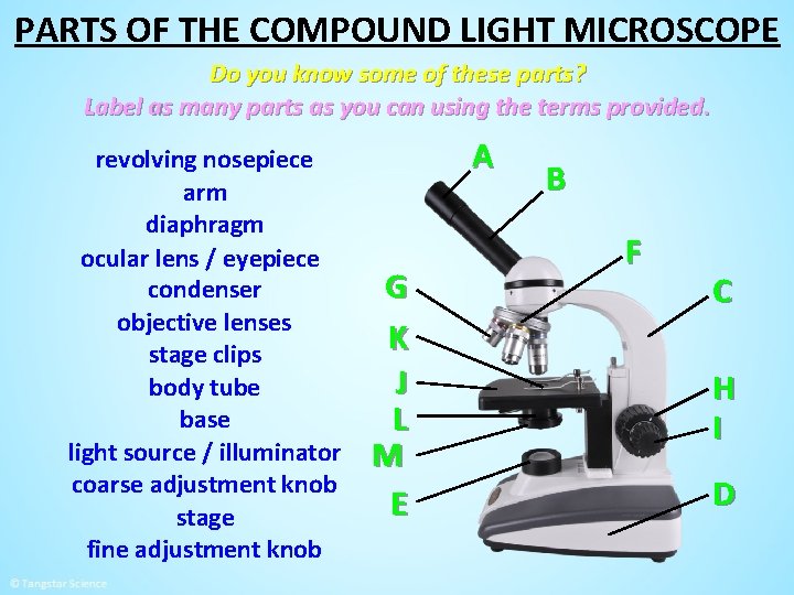
MICROSCOPE PARTS PARTS OF THE COMPOUND LIGHT MICROSCOPE
Electron microscope - Wikipedia An electron microscope is a microscope that uses a beam of accelerated electrons as a source of illumination. As the wavelength of an electron can be up to 100,000 times shorter than that of visible light photons, electron microscopes have a higher resolving power than light microscopes and can reveal the structure of smaller objects.. Electron microscopes use shaped magnetic …

Stereo Microscope PNG - Stereo Microscope Labeled, Stereo ...
Compound Microscope: Definition, Diagram, Parts, Uses, Working ... - BYJUS The parts of a compound microscope can be classified into two: Non-optical parts Optical parts Non-optical parts Base The base is also known as the foot which is either U or horseshoe-shaped. It is a metallic structure that supports the entire microscope. Pillar The connection between the base and the arm are possible through the pillar. Arm

Compound Microscope Parts, Diagram Definition, Application ...
Label a Compound Microscope Diagram | Quizlet Label a Compound Microscope STUDY Learn Flashcards Write Spell Test PLAY Match Gravity Created by Hesi_Study Terms in this set (16) Label this Eyepiece (ocular lens) Label this Body tube Label this Arm Label this Mechanical Stage Control Knobs Label this Coarse Adjustment Knob Label this Fine Adjustment Knob Label this Base Label this

MICROSCOPE Labeling - Part - 3
Fluorescence Microscopy - Explanation and Labelled Images 16.12.2020 · A fluorescence microscope works by combining the magnifying properties of the light microscope with fluorescence emitting properties of compounds. Fluorescence microscopy uses a high-intensity light source that excites a fluorescent molecule called a fluorophore in the sample observed. The samples are labeled with fluorophore where they absorb the high …

Parts of a Compound Microscope and Their Functions
Microscope Types (with labeled diagrams) and Functions Compound microscope labeled diagram. Compound microscope functions: It finds great application in areas of pathology, pedology, forensics etc; Its greater order of magnification allows for deeper study of microbial organisms to Detect the cause of diseases; Study the mineral composition in soils; Examine evidences collected in crime scenes by forensics.

Label the image of a compound light microscope - AnswerPrime.com
Parts of a Compound Microscope and Their Functions - NotesHippo Compound microscope magnification is determined by multiplying the eyepiece and objective powers. When viewed through a 5X eyepiece with a 10X objective, an item is magnified 5 x 10=50 times. The magnification is 10 x 45 = 450 times when using a 10X eyepiece and a 45X objective. How to Use the Compound Microscope
Microscopes Quiz | Biology - Quizizz
10 Best Compound Microscopes (Summer 2022) - The Complete Guide Celestron Labs designed a premium compound microscope with exceptional durability. It comes with a two-year warranty, but it will last longer if you maintain it right. The product includes two eyepieces, a WP-20x and a WF-10x with a pointer. You can change these eyepieces easily to find your perfect adjustment.

Compound Microscope Parts
Microscope Labeling - The Biology Corner Microscope Labeling. Shannan Muskopf May 31, 2018. This simple worksheet pairs with a lesson on the light microscope, where beginning biology students learn the parts of the light microscope and the steps needed to focus a slide under high power. The labeling worksheet could be used as a quiz or as part of direct instruction where students ...

Microscope Parts and Functions
Lab Supplies | Lab Consumables | Lab Equipment | Hurst Scientific We supply lab equipment, lab supplies and lab consumables for most scientific disciplines. Browse our wide range of lab and scientific products online. Delivered Australia wide.

Parts Of A Microscope - Compound Microscope Parts, HD Png ...
Introductory Hydra Activities

Microscopes: A Beginner's Guide

Microscope Terminology

Label the microscope — Science Learning Hub

Compound Microscope Parts, Functions, and Labeled Diagram ...

Compound and Stereo Microscopes | Download Scientific Diagram

What is a Compound Microscope? | Microscope World Blog

Diagram of a Compound Microscope
Monday 10/19/15 AIM: how do the parts of the compound light ...
1.2: Microscopes - Biology LibreTexts

SWIFT SW150 EP1 Compound Microscope of 40X-1000X With 1.3MP ...

Biology label part of microscope

Compound Microscope – Diagram (Parts labelled), Principle and ...

Parts of a microscope with functions and labeled diagram

MICROBIO 16 Parts of a Compound Microscope with Diagram and ...
Label The Parts Of A Compound Microscope Teaching Resources | TpT

Label the microscope — Science Learning Hub
Post a Comment for "43 compound microscope labelling"