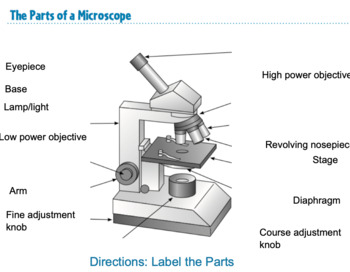
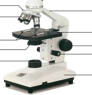
40 label the indicated parts of the microscope.
Cladogram- definition, features, parts, examples (vs Phylogram) Cladogram Definition. A cladogram is the graphical representation of the hypothetical relationship (phylogenetic relationship) between different groups of organisms. It is used in the phylogenetic analysis of organisms to determine the evolutionary relationship between them. The cladogram is derived from Greek words clados and gramma where ... Western Herb Products, Inc. - 615439 - 12/07/2021 | FDA The label indicated that Artemisia absinthium is derived from the leaf part of the plant, however, the specification sheet indicated that the botanical ingredient is derived from the aerial parts ...
(Solved) - (1)Draw a neat diagram of a monocular microscope and label ... transmission microscope ...

Label the indicated parts of the microscope.
5 White Blood Cells Types and Their Functions - New Health Advisor Agranulocytes are free of visible grains under the microscope and include lymphocytes and monocytes. Together, they coordinate with one another to fight off things like cancer, cellular damage, and infectious diseases. Below, detailed information about each type will be discussed. 1. Neutrophils 15 Microscope Parts with Diagram, Location and Function - Study Read A simple microscope has just a lens, stage, and light source. But a compound microscope has many parts like Eyepiece Body Tube Revolving nosepiece Objective lens (Three 10x, 45x, 100x) Coarse adjustment Fine adjustment Arm/handle Rack stop Fixed stage Mechanical Stage Clips Side-ward movement knob Front and back movement knob 8.2: Transmission Electron Microscopy - Chemistry LibreTexts Transmission electron microscopy (TEM) is a form of microscopy which in which a beam of electrons transmits through an extremely thin specimen, and then interacts with the specimen when passing through it. The formation of images in a TEM can be explained by an optical electron beam diagram in Figure 8.2. 1.
Label the indicated parts of the microscope.. Parts of the Microscope with Labeling (also Free Printouts) Let us take a look at the different parts of microscopes and their respective functions. 1. Eyepiece it is the topmost part of the microscope. Through the eyepiece, you can visualize the object being studied. Its magnification capacity ranges between 10 and 15 times. 2. Body tube/Head It is the structure that connects the eyepiece to the lenses. AntiSMASH cluster visualization — MicroScope User Doc v3.16.0 This window allows you to visualize the antiSMASH cluster predictions and its genomic context. The boxes in the middle of the viewer (on the blue line) represent the predicted domains of the predicted secondary metabolite genes. The other boxes in the viewer represent the genomic object existing in the region. Structure of Cell: Definition, Types, Diagram, Functions - Embibe Robert Hooke's discovery was important because it indicated that for the first time living organisms consisted of a number of small structures or units. The ordinary compound microscope of today is a greatly improved design of the original Hooke's microscope. However, the cells which Hooke observed had no information about the organelles ... Neuromuscular junction: Parts, structure and steps | Kenhub The neuromuscular junction: Structure and function. At its simplest, the neuromuscular junction is a type of synapse where neuronal signals from the brain or spinal cord interact with skeletal muscle fibers, causing them to contract. The activation of many muscle fibers together causes muscles to contract, which in turn can produce movement ...
Blood Smear Test: Procedure, Staining & Interpretation Using this method, a mixed drop of blood 1 to 2 mm in diameter is placed in the center line about 1/4 inch from the edge of the microscope slide using a pipette or capillary tube. The slide ... Motility Test (Theory) - Amrita Vishwa Vidyapeetham Virtual Lab A positive result is indicated by diffuse or cloudy growth mostly at the top and bottom of the stab. SIM agar may also be used to detect the presence of H 2 S production. The SIM medium contains peptones and sodium thiosulfate as substrates, and ferrous ammonium sulfate, Fe (NH 4 )SO 4 , as the H 2 S indicator. Researchers boost sensitivity and speed of Raman ... - ScienceDaily Researchers have developed a label-free and non-invasive Raman spectroscopy approach that can acquire microscopic images of biological samples and identify a wide range of biomolecules with... Parts of a microscope with functions and labeled diagram - Microbe Notes Q. List down the 18 parts of a Microscope. 1. Ocular Lens (Eye Piece) 2. Diopter Adjustment 3. Head 4. Nose Piece 5. Objective Lens 6. Arm (Carrying Handle) 7. Mechanical Stage 8. Stage Clip 9. Aperture 10. Diaphragm 11. Condenser 12. Coarse Adjustment 13. Fine Adjustment 14. Illuminator (Light Source) 15. Stage Controls 16. Base 17.
Microscope Quiz: How Much You Know About Microscope Parts ... - ProProfs Projects light upwards through the diaphragm, the specimen, and the lenses. 5. Is used to regulates the amount of light on the specimen. Supports the slide being viewed. Moves the stage up and down for focusing. 6. Is used to support the microscope when carried. Moves the stage slightly to sharpen the image. Thymus: Histology, features, cell types and anatomy | Kenhub Divided into thymic lobules separated by connective tissue septae. Each lobule is made up of a peripheral cortex and an inner medulla. Thymic cortex. Superficial layer: superficial subscapular cells forming a squamous sheath and a blood thymus barrier. Middle layer: stellate thymic epithelial and cytoreticular cells. A. Label the parts of the microscope below. Identify whether the ... answered A. Label the parts of the microscope below. Identify whether the labeled parts are mechanical, magnifying or illuminating parts. Advertisement Answer 5.0 /5 1 norhainefayelatayan Answer: A. Coarse Adjustment Knob B. Fine Adjustment Knob C. Arm D. Head E. Base F. Ocular Lens (Eyepiece) G. Objective H. Stage I. Condenser J. Illuminator Lab Questions Microscope very important that you learn to use the microscope correctly, and can efficiently get images into the proper focus for study Purell Lab #1 - Microscopy-26 Points DUE: Thursday, February 4, 2021 Name Date 1 Click on each of the microscope parts and other items on the lab bench (indicated by question marks) to familiarize yourself with the ...
Marine primary production - Wikipedia
Label-free nanofluidic scattering microscopy of size and mass ... - Nature Label-free methods bypass the following limitations of fluorescence microscopy: (1) attaching a fluorescent label to a target may alter its properties 6,7; (2) there is a limited number of label ...

J* (@Afri_Abana) / Twitter
1.2: Anatomical Position and Planes - Biology LibreTexts Find the indicated structures in the diagrams provided, based on the directional terms given. The structure to find will be one of those at the end of an unlabeled line. A. Label the extensor digitorum (ED) muscle in the figure below. It is: Distal to the anconeus muscle Lateral to the extensor digiti minimi muscle
Microscope Bundle!! - Parts of a Microscope Unit Activities | TpT
Transport of intensity diffraction tomography with non ... - Nature Since its invention in the 1600s, the optical microscope has experienced continuous development and become an indispensable tool for the visualization of micro-scale objects with high resolution in...
Microscope Exercise - Page 1
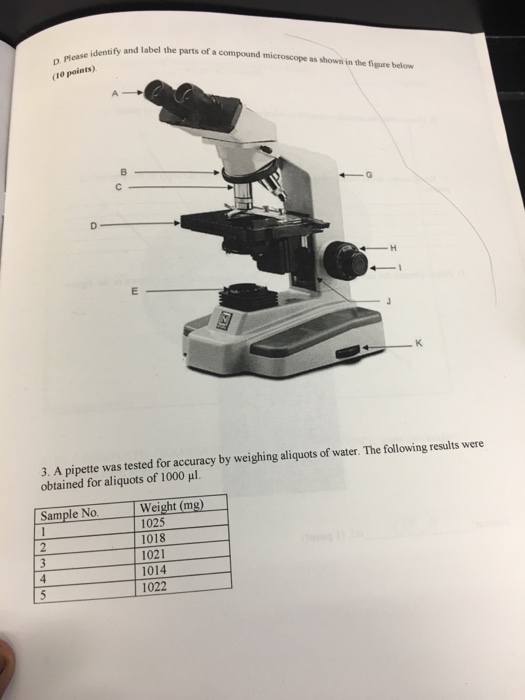
(Get Answer) - Match the step in focusing with the microscope with the ... Care and Structure of the Compound Microscope 1. Label all indicated parts of the microscope. Explain the proper technique for transporting the microscope. ... Parts of a Compound Light Microscope Using the theory found in this lab and the online tutorial in section 2b, match the definitions on the left with the words on the right (not all the

Lab Book - RS 3.pdf - NAME LAB 'I'IME'DATE The Microscope ...
Different Size, Shape and Arrangement of Bacterial Cells Size of Bacterial Cell. The average diameter of spherical bacteria is 0.5-2.0 µm. For rod-shaped or filamentous bacteria, length is 1-10 µm and diameter is 0.25-1 .0 µm. E. coli , a bacillus of about average size is 1.1 to 1.5 µm wide by 2.0 to 6.0 µm long. Spirochaetes occasionally reach 500 µm in length and the cyanobacterium.

Label the microscope — Science Learning Hub
Anatomy, Skin (Integument), Epidermis - StatPearls - NCBI Bookshelf It is made up of three layers, the epidermis, dermis, and the hypodermis, all three of which vary significantly in their anatomy and function. The skin's structure is made up of an intricate network which serves as the body's initial barrier against pathogens, UV light, and chemicals, and mechanical injury.
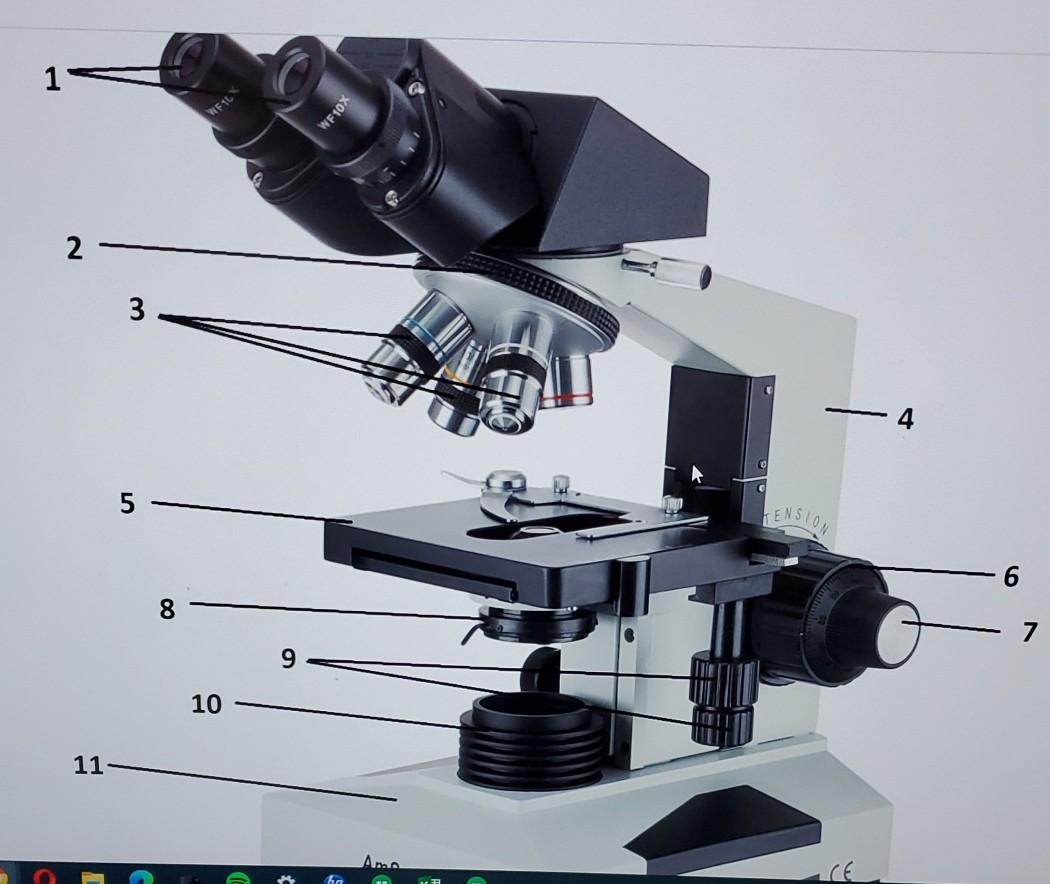
Question: 1 VEL WE10X 2 3 -4 01 TENSION 8 7 9 10 11 Am 12 ...
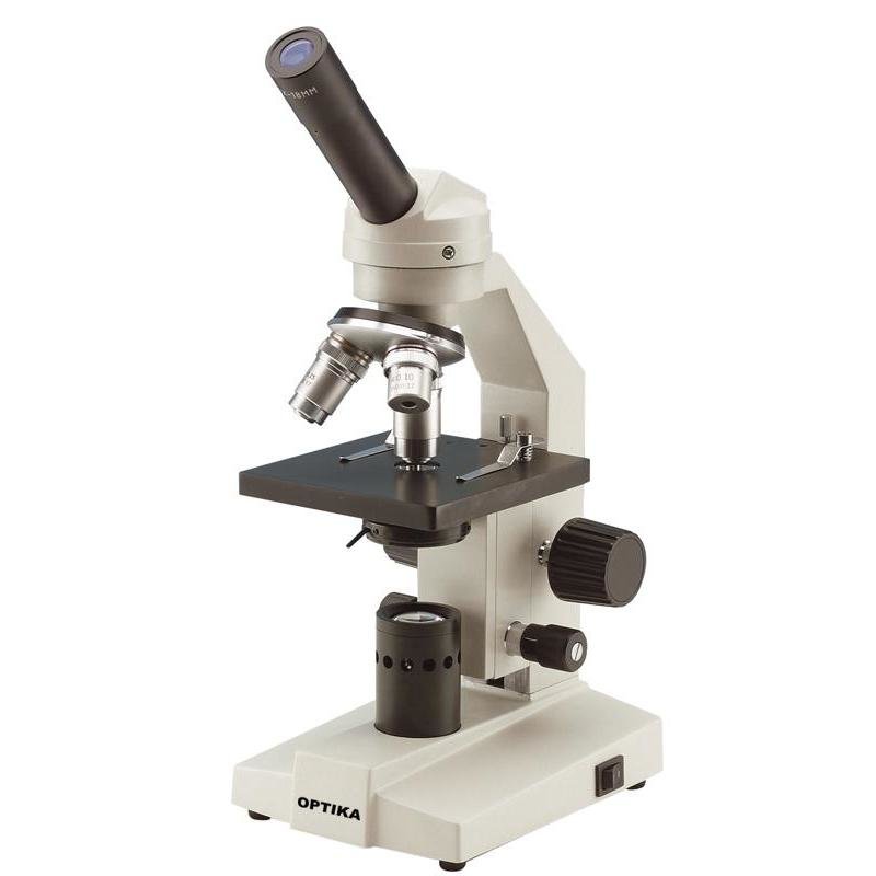
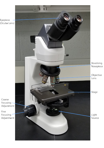
20 most common lab equipment names, pictures and their uses A light microscope placed on a white surface. Photo: pexels.com, @Artem Podrez Source: UGC. A microscope is a popular lab apparatus used to observe things that are too tiny to be observed by the naked human eye. There are many different types of microscopes. A light microscope uses lights and a series of magnifying lenses to observe a tiny ...
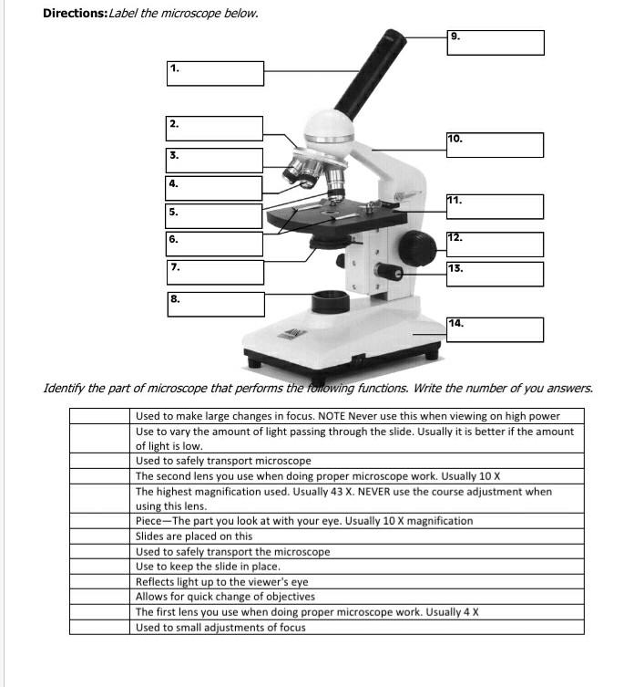
Solved Directions:Label the microscope below. 9. 1. 111 2 ...
Tissue Identification Quiz! - ProProfs Quiz Take up the quiz below and prove it. Questions and Answers 1. A. Stratified squamous B. Simple cuboidal C. Simple columnar 2. A. Cardiac muscle B. Bone C. Skeletal muscle 3. A. Stratified squamous B. Simple columnar C. Simple squamous 4. A. Stratified columnar B. Simple columnar C. Adipose 5. A. Simple cuboidal B. Neuron C. Corn silk 6. A.
bio exercise 3&4 fahmida Usman
Histology, Mast Cells - StatPearls - NCBI Bookshelf Microscopy Light. Mast cells are oval or irregularly shaped cells. Under light microscopy, a dense granular cytoplasm is seen, often obscuring the nucleus and other organelles. When it can be visualized, the nucleus is central, and the cell is mononuclear. Mast cells are found throughout the body in loose connective tissue.
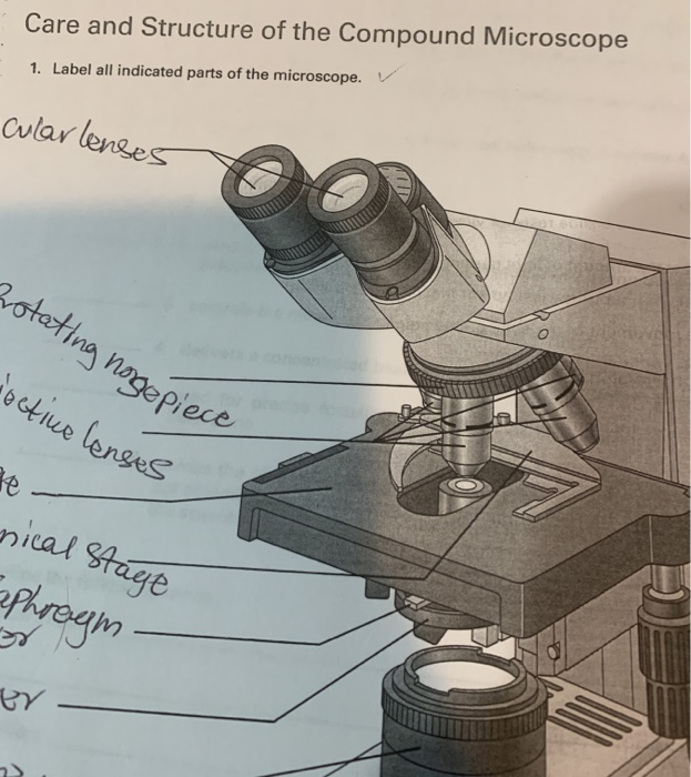
Solved Care and Structure of the Compound Microscope 1 ...
TAGARNO FHD ZAP Digital Video Microscope User Manual Turn the microscope off Press and hold the center button down while turning the power on Keep holding the center button down for 25 seconds Release the center button and turn the power off Turn the power back on and the microscope is set to factory setting 1080P60 ON-SCREEN DISPLAY (OSD)
Microscope Parts Quiz
Cell Organelles- Definition, Structure, Functions, Diagram - Microbe Notes In a plant cell, the cell wall is made up of cellulose, hemicellulose, and proteins while in a fungal cell, it is composed of chitin. A cell wall is multilayered with a middle lamina, a primary cell wall, and a secondary cell wall. The middle lamina contains polysaccharides that provide adhesion and allow binding of the cells to one another.

Microscope Parts & Functions - AmScope
8.2: Transmission Electron Microscopy - Chemistry LibreTexts Transmission electron microscopy (TEM) is a form of microscopy which in which a beam of electrons transmits through an extremely thin specimen, and then interacts with the specimen when passing through it. The formation of images in a TEM can be explained by an optical electron beam diagram in Figure 8.2. 1.
Solved Identify and label the parts of a compound microscope ...
15 Microscope Parts with Diagram, Location and Function - Study Read A simple microscope has just a lens, stage, and light source. But a compound microscope has many parts like Eyepiece Body Tube Revolving nosepiece Objective lens (Three 10x, 45x, 100x) Coarse adjustment Fine adjustment Arm/handle Rack stop Fixed stage Mechanical Stage Clips Side-ward movement knob Front and back movement knob

Bobcat 220 Excavator PDF Service Manual
5 White Blood Cells Types and Their Functions - New Health Advisor Agranulocytes are free of visible grains under the microscope and include lymphocytes and monocytes. Together, they coordinate with one another to fight off things like cancer, cellular damage, and infectious diseases. Below, detailed information about each type will be discussed. 1. Neutrophils

0714nature test worksheet
Parts of a Microscope Quiz
Caterpillar cat d8 l track type tractor dozer bulldozer ...

Compound Microscope Parts – Labeled Diagram and their ...
Special Sense organ

Parts of a Microscope - SmartSchool Systems

Semi automatic Manual Marking Machine YL 360 Sign Nameplate ...
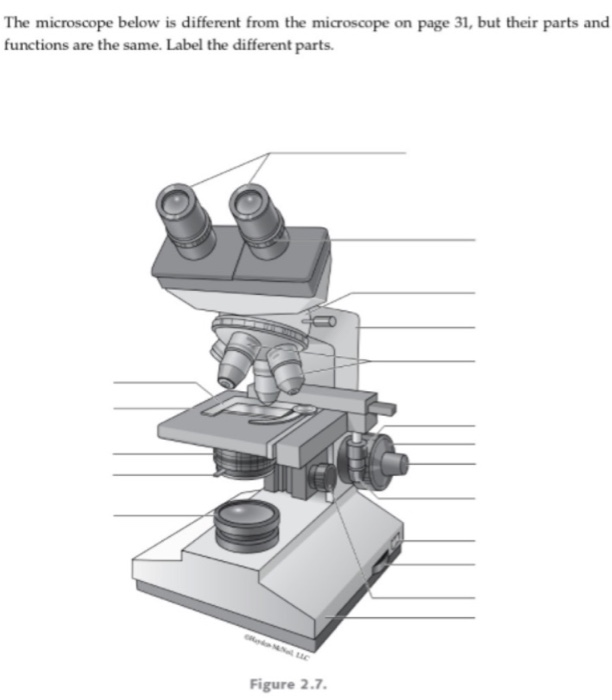
Solved The microscope below is different from the microscope ...
Parts of a microscope with functions and labeled diagram
Untitled
Microscopy and Staining Techniques in Bacteria | Microbiology ...

Labeling the Parts of the Microscope answers.jpg - Eyepiece ...
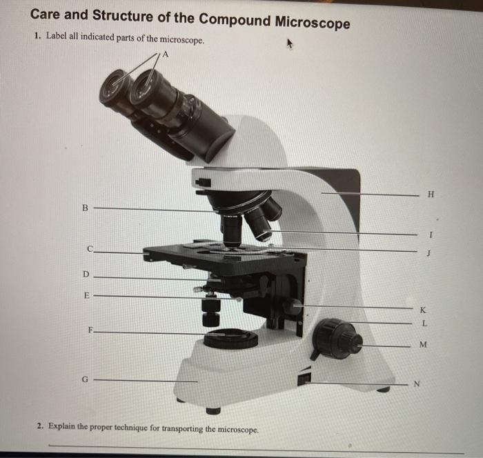
Solved Care and Structure of the Compound Microscope 1 ...
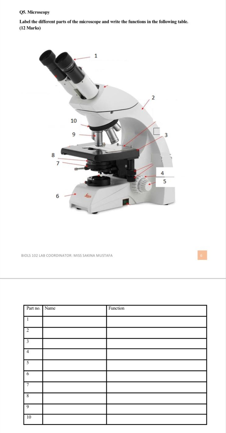
Solved Q5. Microscopy Label the different parts of the ...
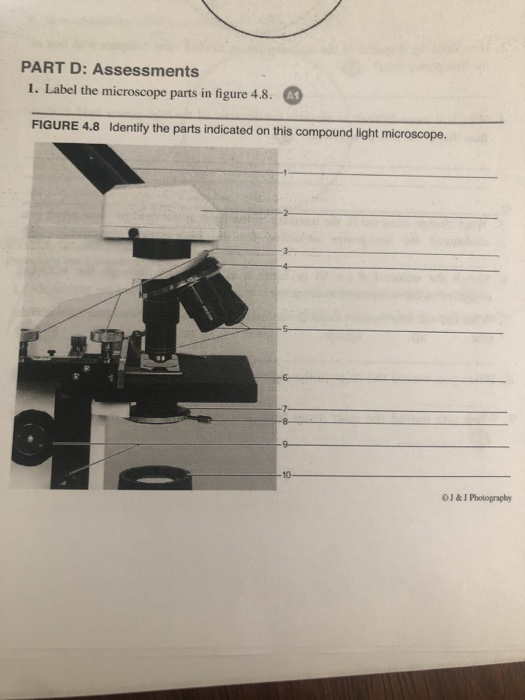
Solved PART D: Assessments 1. Label the microscope parts in ...

Parts of a microscope with functions and labeled diagram
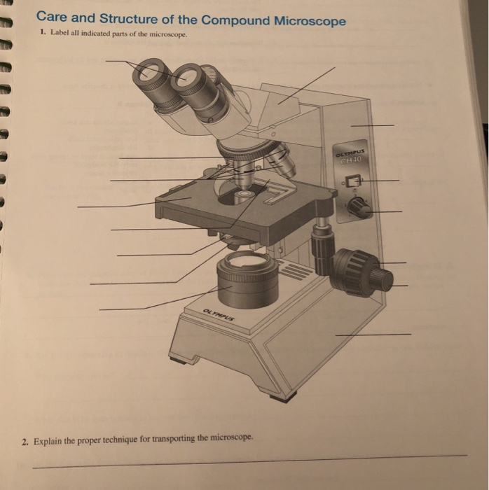
Solved Care and Structure of the Compound Microscope 1 ...
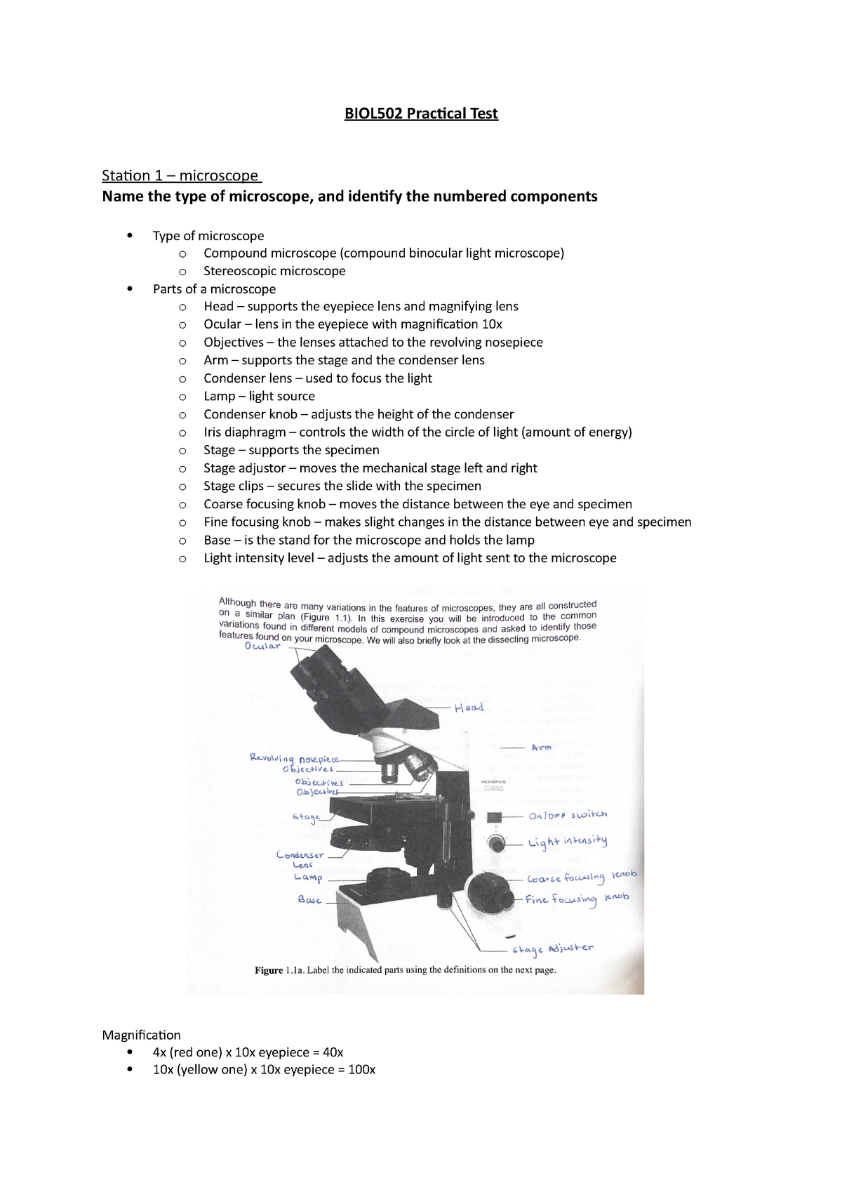
BIOL502 Practical Test - BIOL502 Practical Test Station 1 ...

Compound Microscope Parts – Labeled Diagram and their ...

Compound Microscope Parts – Labeled Diagram and their ...

An Introduction to the Light Microscope, Light Microscopy ...

Review Questions
Solved] Pleas answer all and thanks | Course Hero

Lab Chapter #2 - The Microscope Diagram | Quizlet
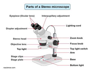
Parts of Stereo Microscope (Dissecting microscope) – labeled ...

Label the microscope — Science Learning Hub
Post a Comment for "40 label the indicated parts of the microscope."