43 eye labelling
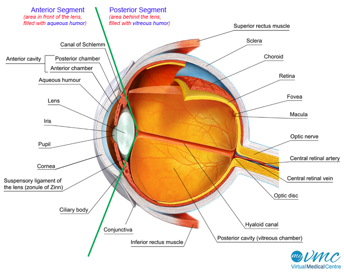
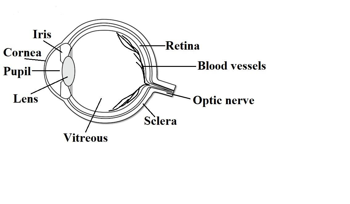
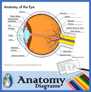
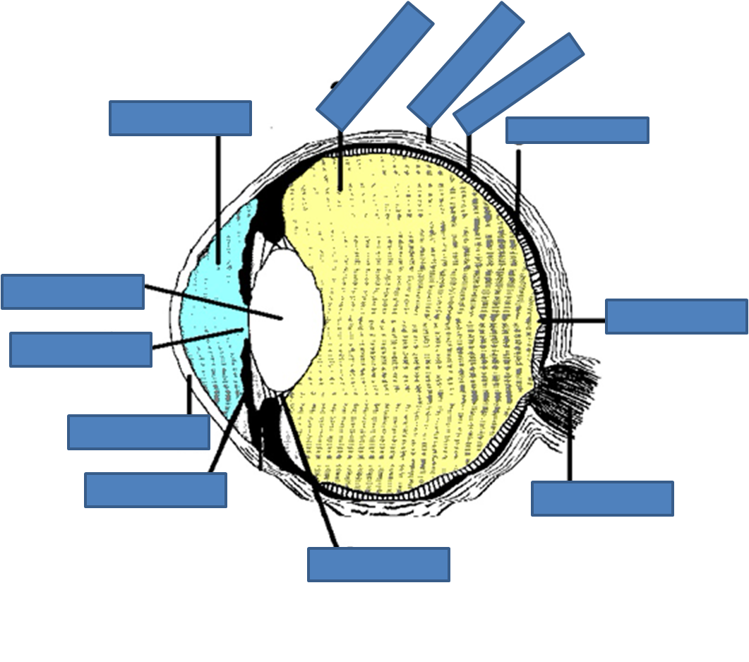
Eye Diagram With Labels and detailed description - BYJUS Well-Labelled Diagram of Eye The anterior chamber of the eye is the space between the cornea and the iris and is filled with a lubricating fluid, aqueous humour. The vascular layer of the eye, known as the choroid contains the connective tissue. The iris and the choroid are connected by the ciliary body. Labelling the eye - Science Learning Hub Labelling the eye Interactive Add to collection Use this interactive to label different parts of the human eye. Drag and drop the text labels onto the boxes next to the diagram. Selecting or hovering over a box will highlight each area in the diagram. Vitrous humour Cornea Lens Schlera Iris Pupil Optic nerve Retina Download Exercise
Eye Anatomy: A Closer Look At the Parts of the Eye - All About Vision When surveyed about the five senses — sight, hearing, taste, smell and touch — people consistently report that their eyesight is the mode of perception they value (and fear losing) most. Despite this, many people don't have a good understanding of the anatomy of the eye, how vision works, and health problems that can affect the eye.
Eye labelling
Anatomy of the Eye | Johns Hopkins Medicine A series of fibers that connects the ciliary body of the eye with the lens, holding it in place. Upper eyelid. Skin that covers the upper part of the eyeball, including the cornea, when closed. Vitreous body. A clear, jelly-like substance that fills the back part of the eye. Find a Treatment Center Diagram of the Eye - Lions Eye Institute The eye - one of the most complex organisms in the human body. It is made up of many different parts working in unison together. In order for the eye to work at its best, all parts must work well collectively. To understand the eye and its functions, it's important to understand how the eye works, see below diagrams for both the external ... Label Parts of the Human Eye Label Parts of the Human Eye Parts of the Eye Select the correct label for each part of the eye. The image is taken from above the left eye. Click on the Score button to see how you did. Incorrect answers will be marked in red.
Eye labelling. Diagram human eye anatomy with label vector image - VectorStock Diagram of human eye anatomy with label illustration. Download a free preview or high-quality Adobe Illustrator (ai), EPS, PDF vectors and high-res JPEG and ... Labeled Eye Diagram - Science Trends The cornea of the eye is composed of five different layers: the corneal epithelium, Bowman's layer, the corneal stroma, Descemet's membrane, and the corneal endothelium. Each of these layers has a function and they work together to transform the light entering the eye as well as protect and support the eye in general. Eye Anatomy: 16 Parts of the Eye & Their Functions - Vision Center The following are parts of the human eyes and their functions: 1. Conjunctiva. The conjunctiva is the membrane covering the sclera (white portion of your eye). The conjunctiva also covers the interior of your eyelids. Conjunctivitis, often known as pink eye, occurs when this thin membrane becomes inflamed or swollen. Labeling of the Eye Quiz - PurposeGames.com Image Quiz Labeling of the Eye by sethf09 2,224 plays 16 questions ~40 sec English 16p 1 too few (you: not rated) Tries 16 [?] Last Played February 22, 2022 - 12:00 am There is a printable worksheet available for download here so you can take the quiz with pen and paper. Remaining 0 Correct 0 Wrong 0 Press play! 0% 08:00.0 Show More
Eye Anatomy: Parts of the Eye and How We See The eye sits in a protective bony socket called the orbit. Six extraocular muscles in the orbit are attached to the eye. These muscles move the eye up and down, side to side, and rotate the eye. The extraocular muscles are attached to the white part of the eye called the sclera. eye labeling Diagram | Quizlet delicate membrane lining the inside of the eyelids and covering the eyeball cornea fibrous transparent layer of clear tissue like a dome that covers the anterior portion of the eyeball (the iris and pupil). It is the first structure to refract (bend) light that enters the eye. sclera Tough white out covering of the eyeball choroid Eye labeling Diagram | Quizlet carries neural impulses from the eye to the brain Iris a ring of muscle tissue that forms the colored portion of the eye around the pupil and controls the size of the pupil opening Cornea The clear tissue that covers the front of the eye Posterior Compartment filled with vitreous humor Pupil opening in the center of the iris Susponsory Ligament Module 1: Labeled Diagram of the Eye - Pinterest Learn about eyes. Which is your dominant eye? Try this to find out. Do a cow eye dissection. And get our free printable eye diagram to label and color.
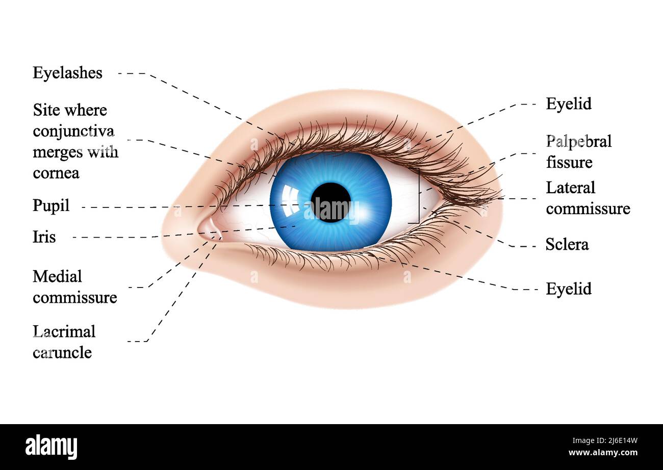
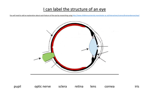
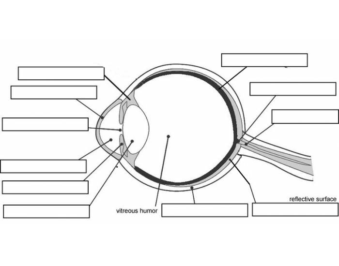
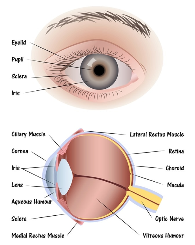
Label the Eye Worksheet - Teacher-Made Learning Resources - Twinkl The first page is a labelling exercise with two diagrams of the human eye. One is a view from the outside, and the other is a more detailed cross-section. Challenge learners to label the parts of the eye diagram. Show more Related Searches PDF Parts of the Eye - National Institutes of Health Eye Diagram Handout Author: National Eye Health Education Program of the National Eye Institute, National Institutes of Health Subject: Handout illustrating parts of the eye Keywords: parts of the eye, eye diagram, vitreous gel, iris, cornea, pupil, lens, optic nerve, macula, retina Created Date: 12/16/2011 12:39:09 PM Labelling the eye - Science Learning Hub Labelling the eye Resource Add to collection The human eye contains structures that allow it to perceive light, movement and colour differences. In this activity, students use online or paper resources to identity and label the main parts of the human eye. By the end of this activity, students should be able to: FREE! - The Human Eye Labeling Activity (Teacher-Made) - Twinkl The first page is a labelling exercise with two diagrams of the human eye. One is a view from the outside, and the other is a more detailed cross-section. Challenge learners to label the parts of the eye diagram. Show more Related Searches
Vision and the eye's anatomy | HealthEngine Blog
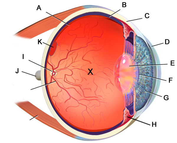
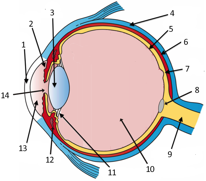
Parts of the human eye - labelling (practice) | Khan Academy Parts of the human eye - labelling Google Classroom The diagram below points to different parts of the human eye. The human eye. Choose the correct labels for the parts shown. Choose all answers that apply: \text A A is the crystalline lens. A \text A A is the crystalline lens. \text B B is the aqueous humour. B \text B B is the aqueous humour.

Draw a diagram of vertical section of human eye and label the ...
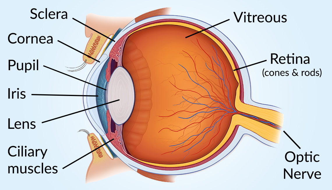
Vision and Eye Diagram: How We See - AARP Cones are responsible for producing the visual sharpness of the eye — seeing road signs when driving, fine print when reading or recognizing facial details like the color of someone's eyes — as well as color vision. "Most of these cones are concentrated in a very specific area in the center of retina, called the macula," says Haugsdal.
Label the Eye
A Picture of the Eye - WebMD Your eye is a slightly asymmetrical globe, about an inch in diameter. The front part (what you see in the mirror) includes: Iris: the colored part Cornea: a clear dome over the iris Pupil: the...
Labelling the eye - Teaching resources
Label the Eye Quiz - PurposeGames Label the Eye by LegoA1 381,527 plays 12 questions ~30 sec English 12p 157 4.26 (you: not rated) Tries Unlimited [?] Last Played February 15, 2023 - 04:09 PM There is a printable worksheet available for download here so you can take the quiz with pen and paper. From the quiz author Title Says It ALL!!!! Remaining 0 Correct 0 Wrong 0 Press play! 0%

Eye diagram to be labeled worksheet
The Eye - Science Quiz The Eye - Science Quiz: Our eyes are highly specialized organs that take in the light reflected off our surroundings and transform it into electrical impulses to send to the brain. The anatomy of the eye is fascinating, and this quiz game will help you memorize the 12 parts of the eye with ease. Light enters our eyes through the pupil, then passes through a lens and the fluid-filled vitreous ...

Saland Vision | Diagram of the Eye | Dallas
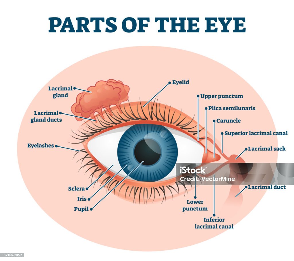
Human Eye Ball Anatomy & Physiology Diagram - eMedicineHealth The orbit is the bony eye socket of the skull. The orbit is formed by the cheekbone, the forehead, the temple, and the side of the nose. The eye is cushioned within the orbit by pads of fat. In addition to the eyeball itself, the orbit contains the muscles that move the eye, blood vessels, and nerves. The orbit also contains the lacrimal gland ...
Human eye anatomy illustration. Parts of the eye, labeled ...
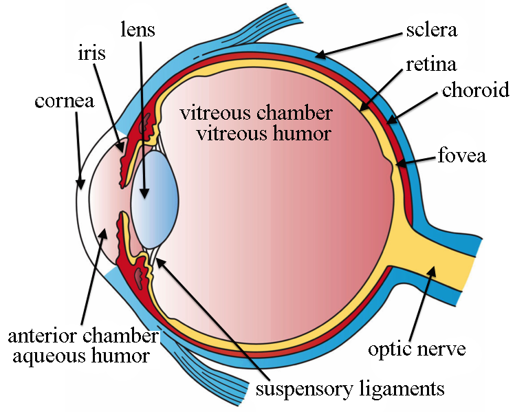
Structure and Functions of Human Eye with labelled Diagram - BYJUS The parts of the eye that are visible externally include the following:- Sclera: It is a white visible portion. It is made up of dense connective tissue and protects the inner parts. Conjunctiva: It lines the sclera and is made up of stratified squamous epithelium.
✓ Solved: Label the parts of the eye:
Eye Diagram Quiz - ProProfs Quiz Try this amazing Eye Diagram Quiz quiz which has been attempted 8049 times by avid quiz takers. Also explore over 71 similar quizzes in this category.
Vision and Eye Diagram: How We See
Quiz: Label The Parts Of The Eye - ProProfs Quiz Take up this quiz and find out how much did you get to understand about the human eye? All the very best to you! Questions and Answers 1. A is pointing to what part of the eye? A. Cornea B. Optic Nerve C. Iris D. Pupil E. Sclera 2. B is pointing to what part of the eye? A. Optic Nerve B. Lens C. Retina D. Pupil E. Iris 3.

Eye Model Labeled - Bing Images | Eye anatomy diagram ...
Human Eye Anatomy Quiz - Sporcle Human Eye Anatomy Can you locate the parts of the human eye? By smac17. Follow. Send a Message. Give Orange. See More by this Creator. Comments. Comments. Bookmark Quiz Bookmark Quiz Bookmark. Favorite. Share with Friends Add To Playlist. Report. View Reports-/5-RATE QUIZ. YOU. MORE INFO. Last Updated. Nov 13, 2017. Last Played.

The diagram shows a cross-section of the human eye. Which ...
Structure and Function of the Eyes - Eye Disorders - MSD Manuals Near the front of the eye, in the area protected by the eyelids, the sclera is covered by a thin, transparent membrane (conjunctiva), which runs to the edge of ...
Draw a labelled sketch of the human eye.
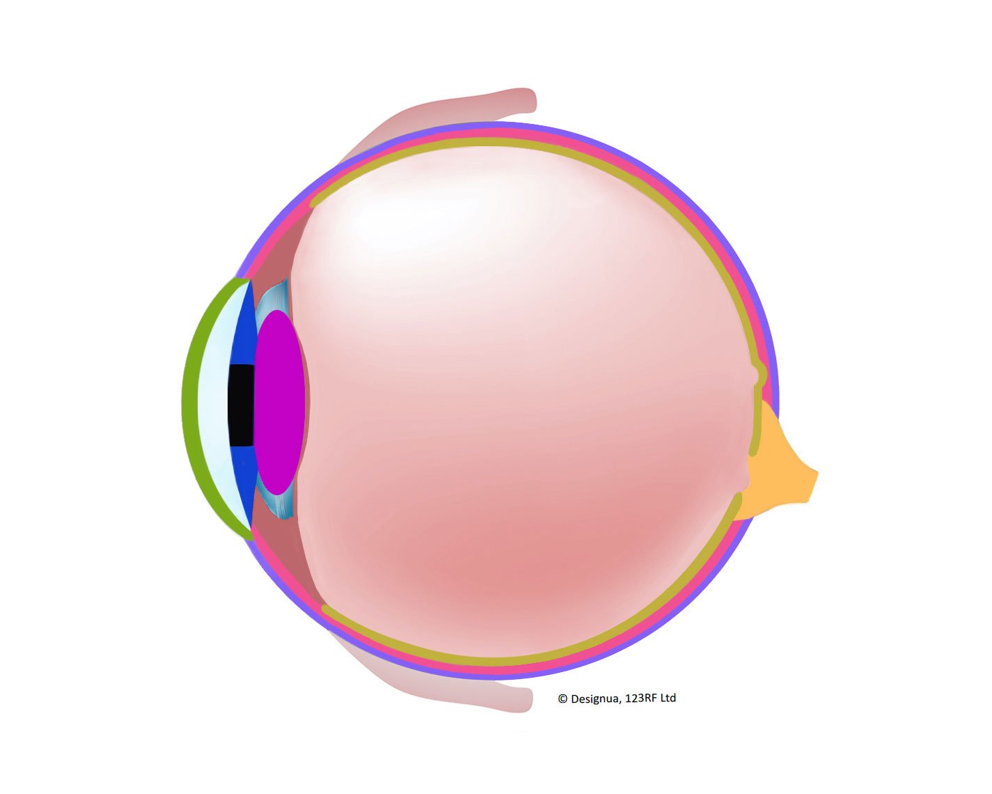
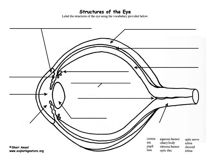
Anatomy of the eye: Quizzes and diagrams | Kenhub Take a look at the diagram of the eyeball above. Here you can see all of the main structures in this area. Spend some time reviewing the name and location of each one, then try to label the eye yourself - without peeking! - using the eye diagram (blank) below. Unlabeled diagram of the eye. Click below to download our free unlabeled diagram of ...

Eye Anatomy with Labeled Structure Scheme for Human Optic ...
Anatomy of the Human Eye - News Medical Retina is the innermost layer of the eyeball structure. Retinal membrane can be imagined as the wall on which the images are projected. The light passing ...
![Cross sectional diagram of human eye [1]. | Download ...](https://www.researchgate.net/publication/276541864/figure/fig1/AS:612895498964992@1523137082339/Cross-sectional-diagram-of-human-eye-1.png)
Cross sectional diagram of human eye [1]. | Download ...
Labelled Diagram of Human Eye, Explanation and Function - VEDANTU Labeled Diagram of Human Eye The eyes of all mammals consist of a non-image-forming photosensitive ganglion within the retina which receives light, adjusts the dimensions of the pupil, regulates the availability of melatonin hormones, and also entertains the body clock.

Label Parts of the Human Eye
Label the Eye - The Biology Corner Label the Eye. Shannan Muskopf December 30, 2019. This worksheet shows an image of the eye with structures numbered. Students practice labeling the eye or teachers can print this to use as an assessment. There are two versions on the google doc and pdf file, one where the word bank is included and another with no word bank for differentiation.

Label the eye parts correctly.i Aqueous humour ii vitreous ...
Label Parts of the Human Eye Label Parts of the Human Eye Parts of the Eye Select the correct label for each part of the eye. The image is taken from above the left eye. Click on the Score button to see how you did. Incorrect answers will be marked in red.
Label the Eye
Diagram of the Eye - Lions Eye Institute The eye - one of the most complex organisms in the human body. It is made up of many different parts working in unison together. In order for the eye to work at its best, all parts must work well collectively. To understand the eye and its functions, it's important to understand how the eye works, see below diagrams for both the external ...
Label Eye Printout - EnchantedLearning.com
Anatomy of the Eye | Johns Hopkins Medicine A series of fibers that connects the ciliary body of the eye with the lens, holding it in place. Upper eyelid. Skin that covers the upper part of the eyeball, including the cornea, when closed. Vitreous body. A clear, jelly-like substance that fills the back part of the eye. Find a Treatment Center

Diagram human eye anatomy with label Royalty Free Vector

Diagram human eye anatomy with label Royalty Free Vector

Eye Model Labeled - Bing Images | Eye anatomy, Eye health ...
What Does the Eye Look Like? – Diagram of the Eye | Harvard ...
Label the eye | Teaching Resources

Complete eye diagram with labels. Courtesy of U.S. National ...
Anatomy of the Eye Diagrams for Coloring/Labeling, with ...

Eye labeling 2 Diagram | Quizlet
Label the Parts of the Eye Quiz
Labelling the eye — Science Learning Hub

Eye Diagram Quiz - ProProfs Quiz

a Draw a labelled diagram of the human eye. Label the ...
Parts Of The Eye Labeled Vector Illustration Diagram Stock ...

Diagram human eye anatomy with label Royalty Free Vector
:max_bytes(150000):strip_icc()/GettyImages-695204442-b9320f82932c49bcac765167b95f4af6.jpg)
Structure and Function of the Human Eye
Anatomy of the Human Eye

Twitter post- Muscles of the eye labelling activity | Arizona ...

Photograph | Eye Anatomy (labeled), illustration | Science ...
Eye Structure Labeling with Vocabulary List
Eye Anatomy Diagram - EnchantedLearning.com
Eye labelling quiz. | Quiz

Diagram of human eye anatomy with label 1928861 Vector Art at ...

2,424 Eye Anatomy Labeling Images, Stock Photos & Vectors ...
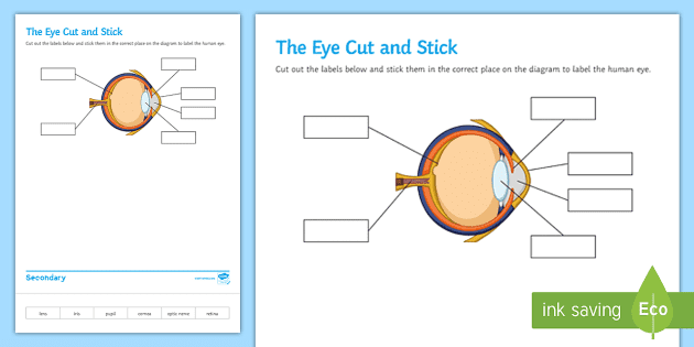
FREE! - The Human Eye Labeling Activity (Teacher-Made)

Human Eye – Origami Organelles
Post a Comment for "43 eye labelling"